The discovery of a circulatory system called the interstitium that operates “in between” the lymphatic and cardiovascular systems has profound implications for the diagnosis and treatment of many conditions—and may forge new links between Western, Eastern, and other traditional medicines.
By Mary Ann Meyers
Rebecca Wells grew up in New England—in Northampton, Massachusetts, a town that lies alongside the Connecticut River; a few miles north in the village of Millers Falls, named for the adjacent river; and in Cumberland, Rhode Island, on the banks of the Blackstone River. The waterways of her childhood and the Quinnipiac River running through New Haven, where she lived as a young adult, were once significant for transport, trade routes carrying canoes of American Indians for centuries before European colonization.
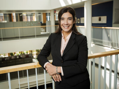
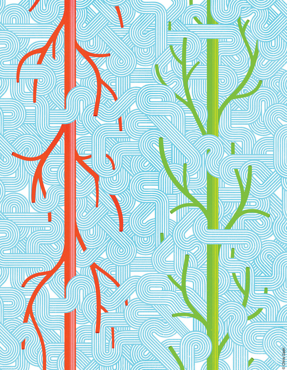
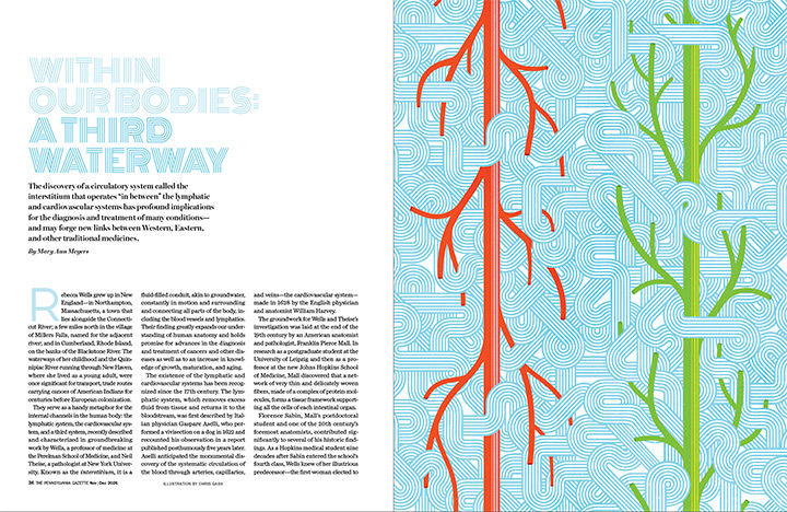
They serve as a handy metaphor for the internal channels in the human body: the lymphatic system, the cardiovascular system, and a third system, recently described and characterized in groundbreaking work by Wells, a professor of medicine at the Perelman School of Medicine, and Neil Theise, a pathologist at New York University. Known as the interstitium, it is a fluid-filled conduit, akin to groundwater, constantly in motion and surrounding and connecting all parts of the body, including the blood vessels and lymphatics. Their finding greatly expands our understanding of human anatomy and holds promise for advances in the diagnosis and treatment of cancers and other diseases as well as to an increase in knowledge of growth, maturation, and aging.
The existence of the lymphatic and cardiovascular systems has been recognized since the 17th century. The lymphatic system, which removes excess fluid from tissue and returns it to the bloodstream, was first described by Italian physician Gaspare Aselli, who performed a vivisection on a dog in 1622 and recounted his observation in a report published posthumously five years later. Aselli anticipated the monumental discovery of the systematic circulation of the blood through arteries, capillaries, and veins—the cardiovascular system—made in 1628 by the English physician and anatomist William Harvey.
The groundwork for Wells and Theise’s investigation was laid at the end of the 19th century by an American anatomist and pathologist, Franklin Pierce Mall. In research as a postgraduate student at the University of Leipzig and then as a professor at the new Johns Hopkins School of Medicine, Mall discovered that a network of very thin and delicately woven fibers, made of a complex of protein molecules, forms a tissue framework supporting all the cells of each intestinal organ.
Florence Sabin, Mall’s postdoctoral student and one of the 20th century’s foremost anatomists, contributed significantly to several of his historic findings. As a Hopkins medical student nine decades after Sabin entered the school’s fourth class, Wells knew of her illustrious predecessor—the first woman elected to the National Academy of Sciences—but years would pass before she drew on Mall’s work in her own collaboration with Theise.
Wells had majored in molecular biophysics and biochemistry at Yale, graduating magna cum laude in 1983, and chose Hopkins for medical school because, as she put it, “it seemed like the right fit.” Her residency at Brigham and Women’s Hospital in Boston was followed by an eight-year stretch of research and clinical fellowships, first in nephrology and then in gastroenterology, at Brigham and at Harvard Medical School. For three years during this period, she was also a visiting scientist at the Whitehead Institute, considered the world’s leading institution for research in molecular biology and genetics.
In laboratories at these institutions, Wells took a deep dive into the complexities of several proteins, the macromolecules that build, repair, and regulate nearly every system in the human body, supporting structure, metabolism, immunity, and communication. Her papers, now numbering more than 150, detailed her investigations of a surface protein found in many mammal tissues that plays a role in cell activation and growth; another protein’s involvement in the movement of amino acids across cell membranes and its role in human inherited disease; and the chromosomal location of a gene expressed primarily in the segment of the kidneys responsible for reabsorbing glucose in the bloodstream.
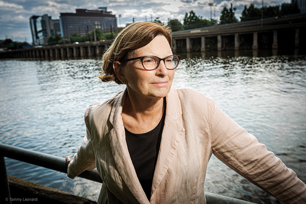
Wells next spent four years on the faculty of Yale Medical School before joining the Perelman School faculty as an assistant professor in 2002. Promoted to full professor in 2017, she became vice chief for research in the Division of Gastroenterology and Hepatology two years later. In 2021 she was named codirector of the Center for Engineering MechanoBiology, a National Science Foundation–funded center with the goal of developing a research and educational program integrating concepts in the physical sciences, engineering, and biology.
Wells’s lab studies the intersection between the extracellular matrix (the scaffolding that holds tissues together) and the response of tissues to physical force in the context of wound healing, using the liver and biliary tree (a branching network of bile ducts connected to the liver, gallbladder, and small intestine) as model tissues. This work led to an ongoing project examining the mechanics of the scarred (fibrotic and cirrhotic) liver and the impact of scarring on the development of liver cancer, as well as projects on the pediatric disease biliary atresia. Her most cited article focuses on the resistance to deformation (mechanical rigidity) of extracellular tissues, a factor that influences cell behavior and disease progression. Wells describes the goal of her research as developing “a unified and comprehensive model of liver and biliary fibrosis”—the gradual scarring of these tissues caused by long-term injury.
Wells and Theise first met when she was a junior faculty member at Yale and Theise was on sabbatical from NYU working in the lab next to hers. They became friends with shared professional interests, but it wasn’t until 2016 that they began to collaborate. Theise and a colleague, David Carr-Locke (also a former colleague of Wells), were exploring an unusual structure Carr-Locke observed in the bile duct. A leader in the field of therapeutic endoscopy who is now professor of medicine at Weill Cornell Medicine, Carr-Locke had used confocal endomicroscopy, a powerful new technique for examining real-time microscopic and histological features inside the body that combines the benefits of endoscopy (a procedure to examine internal bodily structures at close range) with that of laser microscopy.
Theise had questions about the fluid-filled structure he thought he observed, and asked Wells for help. He brought some slides to her lab at Penn for her group to examine using yet another microscopy technique that produces high-resolution images of biological tissues without the need for fluorescent dyes or stains. Wells says she still remembers the two of them sitting in a completely dark room with Gordon Ruthel, the technical director of the Penn School of Veterinary Medicine’s microscopy core, and cheering when the images came on the screen showing the detailed architecture of collagen in tiny canals that connect some of the organs in the digestive system.
Wells and Theise first detailed their discovery in a 2018 article in Nature. The two scientists, along with Carr-Locke, gastroenterologist Petros Benias (co-chief of endoscopy at the Robert Wood Johnson Medical School), and other colleagues reported on the structure and distribution of a hitherto unrecognized channel in human tissues that appeared to be a widespread, fluid-filled network of collagen and elastin fibers—proteins that work together to provide strength, support, and elasticity to tissues throughout the body.
Using confocal endomicroscopy to enhance resolution and contrast, they observed a previously underappreciated fine mesh of fibers in sinuses, the digestive and urinary systems, the skin’s middle layer, and the supportive soft tissues around major airways leading to the lungs, blood vessels, and muscles. They dubbed it the interstitium because it’s between things—a layer between the surface and something deeper. In essence, what they saw, as Wells put it, was a “fluid-filled highway with significant roles in lymph production (the fluid flowing through interstitial spaces enters lymphatic vessels of the lymph system) and potential impacts on cancer, edema, and fibrosis.”
The natural movements of the body—the pulsing of heart and blood vessels, the rhythmic squeezing that moves food through the digestive tract, the flexing of muscles—appeared to push fluid through the network of spaces supported by a lattice of connective tissue proteins. Their initial study raised for its authors a multitude of questions: What were the characteristics and drivers of the fluid flow? Were signals emitted during the process? Did the interstitium, reactively distensible or compressible, act as a shock absorber protecting tissue from stress-related damage? How and why do cancer cells spread through these spaces? Could direct sampling of the interstitial fluid be a diagnostic tool?
At the least, their findings necessitated reconsideration of many of the normal functional activities of different organs and of disordered fluid dynamics in the setting of disease. Most importantly, their hypothesis was that the pattern they observed, with 21st century tools unavailable to Mall and Sabin, suggested that widespread layers of the body thought to be solid connective tissues instead contained fluid-filled spaces, comprising a previously unknown third fluid-filled system in the body. But more work was needed to show that these spaces were continuous throughout the body.
Tattoos gave Wells and Theise the experimental evidence they needed to confirm that the interstitial spaces in the human body are unbroken networks across tissues and organs. Their landmark study published in Communications Biology, an open-access journal of Nature, described research using human tissue samples that showed non-biological tracers (tattoo ink and silver) injected into the interstitial spaces of the colon or applied to the skin penetrated deeper than expected and could be tracked into adjacent tissue compartments and tissue, indicating connectivity. Wells’s team had previously identified hyaluronic acid, a large and water-associated chain of sugar molecules that is a major component of the matrix outside of cells, in interstitial spaces. Theise was able to have tissues stained to show hyaluronic acid connections within and between tissues and surrounding nerves and vessels, confirming the presence of a continuous fluid-filled matrix.
The reason no one had seen these spaces before, Wells and Theise agree, is because of the common practice in medicine of examining “fixed” tissue, meaning that when tissue is prepared for microscopic viewing, it has been treated with chemicals and dehydrated, collapsing the connective protein meshwork surrounding once fluid-filled compartments of tissue and making them appear solid, an assumption about the nature of human anatomy that held for decades. Looking at live tissue with confocal endomicroscopy upended the old view. Wells, Theise, and their colleagues recognized that the fluid-filled spaces in living tissue corresponded with “cracks” seen in the solid areas of fixed tissue sections on microscope slides.
It led to what Wells called “the recognition of a continuous third system throughout the body”—a previously unmapped waterway, perhaps more comparable to the sponge-like system of groundwater than a river but clearly connected to the river-like networks of blood vasculature and lymphatics. The interstitium wasn’t passive, simply holding tissues together; rather, as Wells said, “it guided cells into their proper anatomic organization,” serving as a vast communication network in the body that is four times the size of the cardiovascular system in terms of fluid flow.
The implications of the discovery of the previously unknown feature of human anatomy made by Wells and her colleagues are potentially profound. Their findings could increase our understanding of a variety of conditions and diseases, from how scars form after injury to what triggers some aspects of Crohn’s disease to how certain cancers and infections spread. Further studies are needed to determine the details of the interstitial route taken by some migrating tumor cells as they metastasize to distant parts of the body.
Because of its pervasiveness, Wells knows that the interstitium is “a necessary compartment through which cells enter and exit nearly all tissues” in many human malignancies. She and others are developing ways of determining how malignant and infectious diseases might alter interstitial spaces, including what molecular signals might be sent along the internal pathway, which Wells has come to see as a “signaling hub,” and the means of blocking those that lead inevitably to further tissue and organ damage. Wells is hopeful that further research on the interstitial network will lead to a better understanding of disease trajectories but also to a better understanding of human development and aging.
The developmental origins of the interstitial spaces are not fully understood, but the pathways they form are one of the earliest structural developments in the body. Moreover, the fact that simple multicellular organisms—for example, the jellyfish-like hydra—have anatomical structures with similarities to the interstitium, Wells notes, “is consistent with the interstitium being the most primitive of the three circulatory networks.” It probably arose early in the evolution of animals. As she and Theise wrote in a recent article in the Annual Review of Cell and Developmental Biology, “function as a multicellular organism, not just an aggregate of cells, requires an extracellular means of transmitting intercellular signals.” Given the resemblance of mammalian interstitial spaces to continuous extracellular spaces in plant tissues, including plant cell walls, moreover, they suggested that the latter might have arisen by convergent evolution whereby unrelated species evolve similar traits or adaptations in response to similar environmental pressures.
As a matter of human cultural evolution, particularly cross-cultural exchange, the researchers are intrigued by the possible link that their description of the interstitium suggests between traditional and nontraditional Western medicine and, especially, between Western and Eastern medicine. The worldwide attention paid to the papers Wells and her collaborators have published on their ongoing studies is opening up a more extensive conversation about the connections between Eastern and Western understandings of physiology.
Wells notes that amidst all the accolades they received from their scientific peers were “observations from some in the osteopathic and integrative health communities that their findings weren’t really so novel after all, as the interstitial network was simply a concept not captured in allopathic medicine.” These views “were not far off the mark,” she says, recalling that “in medical school, we threw away connective tissue as not worth our attention. It was considered a biological byproduct. Modern medicine is guided by cell doctrine,” she explains. The theory is a foundational principle in biology stating that all living organisms are composed of cells, that the cell is the basic structural and functional unit of life, and that all cells arise from preexisting cells. “It left little room for the idea that there might be a continuous and multiscale network of open channels filled with fluid,” Wells says.
But prior to the invention of the microscope in the mid-17th century, there was a debate that went back to Greek philosophy and medicine about how the body’s parts were organized and how they functioned together that included hypothesized intermediate spaces or regions that are perhaps best understood as a concept of “between-ness.” Physicians associated with the medical writing attributed to Hippocrates generally understood the body as a network—cavities, channels, pores, membranes, vessels—facilitating the movement of air, blood, phlegm, bile, or other humors. These spaces were hidden but not fully empty; rather, defined by their contents, and in the view of Greek physicians, integral to the body’s mechanics and physiology. Surviving fragments of the fifth century BCE works of Alcmaeon of Croton, the first recorded practitioner of human dissection and one of the earliest figures to integrate natural philosophy with medical practice, helped establish the idea that anatomical function depends upon connections through intermediate pathways. Plato presents the body as intrinsically porous, and his famous and enigmatic concept of chōra is viewed by many modern scholars not as a receptacle but as a kind of “generative between” through which everything comes into existence. Not merely anatomical features, the intermediate spaces provided a key to both a medical and a philosophical understanding of the human form and how the body functioned as a living and unified whole.
In our own day, osteopathic philosophy and practice are based on the idea of communication between bodily systems and the interrelationship of structure and function. Osteopathy not only emphasizes the body’s self-healing capacity but also the interconnection of body systems. Osteopaths recognize that fasciae (one part of the interstitial system) not only delineate anatomic features but believe they simultaneously connect the entire organism into one functional unit.
But whatever the discovery of the interstitium may come to suggest about a possible new linkage between the allopathic and osteopathic schools of Western evidence-based medicine, the correlates to the ancient Chinese practice of acupuncture are provoking widespread interest. Wells notes that “the response to our work of some experts in Chinese medicine, was to shout: ‘That’s what we have been talking about!’”
Most historians believe acupuncture originated in China about 3,000 years ago. It spread to Korea in the sixth century, then to Japan, and later to Europe and the United States. In the 20th century, as acupuncture was gaining acceptance in Western countries, practitioners sometimes set aside spiritual elements that appeared to conflict with scientific knowledge and focused solely on the practice of inserting one or more thin needles into specific sites on the body surface for therapeutic purposes. Although most commonly used for pain relief, acupuncture may also be applied to treat a range of conditions, generally alongside other therapies.
Early acupuncture theory drew on concepts central to traditional Chinese medicine (TCM), such as qi, usually translated as ‘energy’. Qi was believed to flow from the body’s primary organs to the “superficial” tissues—the skin, muscles, tendons, bones, and joints—through channels called meridians. Wells cites a study in which fluorescein dye injected into the forearms of volunteers at an acupoint “tracked along the meridian lines in a pattern that,” she says, “maps very well to the interstitium.”
Chinese scientists have suggested other ways in which traditional Chinese medicine may correspond to the interstitial system. One hypothesis is that the classical TCM concept of the “Sanjiao-Mocou” system is analogous. In TCM theory, the enigmatic tri-part San Jiao (Triple Energizer or Triple Burner) is regarded as the largest of the six Fu-organs, all of which are considered yang organs, meaning they are hollow and primarily involved in the transmission, transformation, and excretion of food, fluids, and waste. The San Jiao has been related to the interstitium of the visceral organs in terms of both its structure and function, according to Wells, and possibly even its developmental origin. It is considered a “waterway” like the interstitial channels. It is thought to have the ability to influence other organs and overall health, mainly through the free movement of qi. As scientists at China Medical University in Shenyang wrote in Frontiers in Physiology: “The very fluidity and openness of these contemporary anatomical models—their departure from rigid, preexisting categorical boundaries—may make them well suited to serve as a conceptual bridge between Western anatomical science and the holistic, systems-based framework of TCM.”
Skilled people involved in shamanistic healing practices and aboriginal traditional medicine also recognize an affinity with their own ways of understanding the body when the interstitium is described to them. “The links are speculative,” Wells says, “and we need to learn more from those who have non-Western perspectives on healing. But I like to think we are moving toward a more global way of interpreting what we are doing as scientists exploring human anatomy. Maybe our work can create a kind of cultural interstitium.”
In any case, it is forcing Wells and her colleagues to reevaluate and integrate existing data on many aspects of anatomy and their relevance to health and disease, including metastasis. High on the list of new questions for which they are seeking answers are ones about fluid flow. The time scale and distance of fluid movement in interstitial spaces, its velocity and diffusion, could be relevant to cell behavior, including of cancerous cells that evade normal regulatory signals. How, Wells asks, does interstitial flow connect to and interact with lymphatic and blood circulation? Will learning the details enable us to detect cancer and other diseases early and maybe predict their course?
Despite the immensity of scientific knowledge that exists about our physical selves, the picture remains profoundly incomplete. Wells is trying to add detail about structure and function—knowledge of human bodies that are, as the awe-struck psalmist wrote long ago, so “fearfully and wonderfully made.” As we have learned over and over again, it is basic biomedical research, the relentless search to fill the gaps in our understanding, combined with clinical observation, that has led time and again to future cures. From Old Chatham, New York, a hamlet in Columbia County between Kinderhook Creek and Stony Kill, Wells commutes to her lab at Penn, a few blocks from the Schuylkill River, to explore the intricate design and dynamic processes of the anatomical systems that are essential to life and health.
Mary Ann Meyers Gr’76 is a longtime contributor to the Gazette.