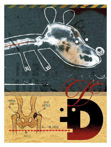
Illustration ©Walter Vasconcelos
Class of ’70 | For Dr. Gail Smith MtE’70 V’74 Gr’82, who recently received the prestigious Iams Saki Paatsama Award from the World Small Animal Veterinary Association, it was his early training in materials science and engineering, coupled with his vet-school training in canine anatomy, that allowed him to “unravel some of the mechanical mysteries of the hip,” in his words. That training led to the development of a pioneering radiographic technique for diagnosing canine hip dysplasia: PennHIP, whose last three letters stand for Hip Improvement Program. (Smith is professor of veterinary orthopedic surgery and chair of clinical studies at the School of Veterinary Medicine, where the program was developed—hence the Penn part of the name.)
What PennHIP does is measure, with considerable precision, the amount of joint laxity, or looseness, in the hips. From that, a vet or breeder can predict the odds and potential severity of the hip dysplasia—defined as abnormal or faulty development of the hip—which often leads to degenerative joint disease or osteoarthritis. Considering that hip dysplasia afflicts more than 50 percent of such popular breeds as golden retrievers, Labrador retrievers, and German shepherds, PennHIP is a very important diagnostic tool.
When Smith began looking at the problem as a junior faculty member, vets were taking x-rays of dogs in the “frontal plane position,” with rear legs extended. It was essentially the same position used to x-ray the hips of humans, and though it was effective for people, it was inconclusive for dogs.
Smith and his colleagues supplemented that angle with two new positions: the “compression view” (with the femoral head pushed in toward the hip socket) and the “distraction view” (with the femoral head pulled away from the socket). The distraction view in particular allows the PennHIP Analysis Center to measure, in millimeters, the amount of play between the femur and socket.
Once they had fine-tuned the technique and proved the correlation between hip laxity and dysplasia, they teamed up with International Canine Genetics, which was later bought by Symbiotics, to market PennHIP. He has since “brought it back to Penn” as a not-for-profit program (www.pennhip.org). In addition to the diagnostic technique itself, PennHIP is a network of trained veterinarians and a medical database for scientific analysis.
So far, some 1,400 vets have been trained to perform the PennHIP radiographic procedure. Yet the number of vets who use the old, conventional system, supported by the Orthopedic Foundation for Animals (OFA), is even greater.
“I’m fighting the old paradigm on hip evaluation, and that’s a huge political mass and mess—our science is convincing and eventually I am confident PennHIP will prevail,” says Smith. “We now do 8,000 dogs a year, and the OFA, our prime competitor, does 50,000. We would like to get our numbers much higher. We’re trying to get corporate support for educating veterinarians, breeders, and pet owners to use this technology.”
His advice to breeders? “Tighter hips are better hips.” If detected early, an owner can address the dog’s condition through diet and exercise. “It’s a matter of educating the world,” he adds. “This method allows the dog owners, not just the breeder, to know if the dog is at risk for osteoarthritis. And now we have some strategies to offset that genetic risk.”
The most important one is “keeping your dog thin,” notes Smith, who recently completed a study that shows that keeping dogs thin helps them live longer and “offsets the genetic predisposition to hip dysplasia.”
Winning the Iams Saki Paatsama Award was a “recognition by the scientific community that your science is good and powerful,” Smith says. “Eventually the overthrow of the old method will take place. It’s just a matter of time.” —S.H.


