
A dog’s brain surgery could have implications for human care.
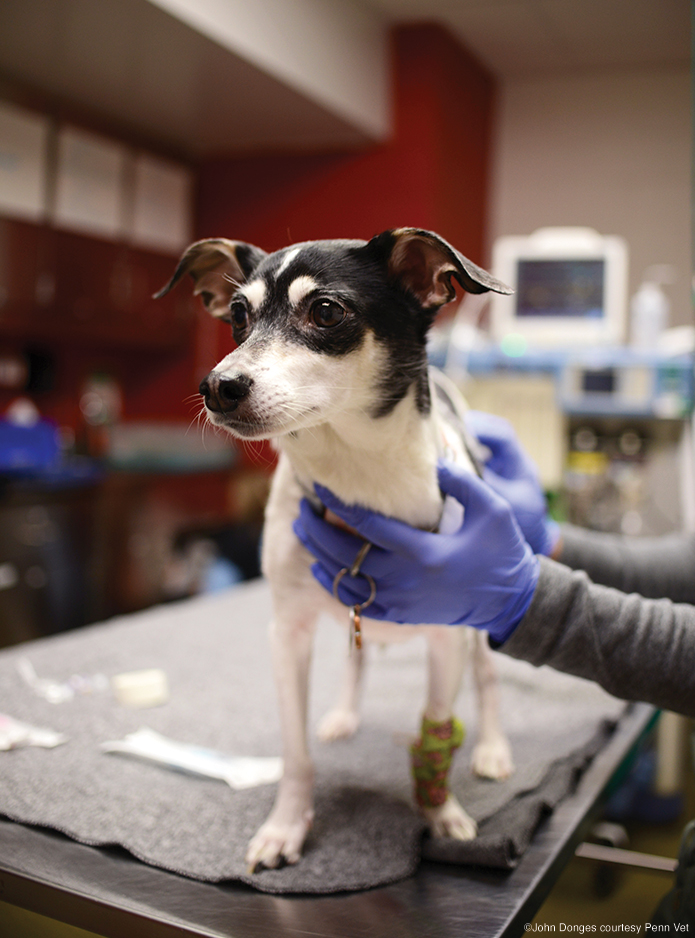
Geddy, a nine-year-old mixed breed 18-pound terrier, is no stranger to drama. Her original owner purchased the pup as a gift for his girlfriend. One small problem: he was driving a stolen car. When the police tried to pull him over, a chase ensued and, as Geddy’s current owner, Michael Crotty, tells it, “he left Geddy in the car and ran.” With the car thief in custody, the police took the abandoned puppy to a local veterinarian because there was no animal shelter in the area. Eventually Geddy was listed on the pet adoption website Petfinder, where Crotty found her, and the tiny pup found her forever home in Mechanicsburg, Pennsylvania, with Crotty, his wife Erica, and their other dog, a rat terrier named Lemmy.
Everything was great until August of last year, when Geddy had a series of violent seizures two nights in a row. The local vet recommended seeing a neurologist, and the Crottys chose to bring Geddy to Penn Vet’s Ryan Veterinary Hospital.
And so began Geddy’s dramatic second act.
On initial examination, Penn Vet doctors Tessa Arendt, specialty intern in neurology, and Wojciech Panek, assistant professor of neurology and neurosurgery in the Department of Clinical Sciences and Advanced Medicine, were prioritizing neoplasia, an abnormal growth of cells. Geddy’s MRI revealed a large lesion in the forebrain or frontal lobe of her brain that, on the imaging, looked like glioma or glioblastoma, a deadly form of brain cancer. A biopsy was the only way to know for sure, but from the size of the mass, they suspected Geddy had about two to three months to live. Since the tumor was at the front of her head, surgery was an expensive possibility, but there was no guarantee they could remove all the mass. Crotty didn’t hesitate. “We had nothing to lose with two to three months,” he recalls. “Let’s do everything we can. Pull out all the stops.”
A few weeks after being diagnosed, Geddy was on her way to making medical history.
Not all owners are able to justify, financially or psychologically, a stop-at-nothing treatment plan. But in addition to potentially lengthening Geddy’s life, Panek knew the surgery would be an opportunity to pioneer a new procedure that might one day provide a more cost-effective means of treating other cases like hers. The centerpiece technique in Geddy’s surgery was use of an augmented-reality surgery system, Visar, that projects the patient’s three-dimensional scans onto the operative field. That meant there was no need to anesthetize Geddy for a second MRI to pinpoint the exact location of the tumor with markers or fiducials. The system was developed by a company called Novarad, of which Wendell Gibby GM’88 is the founder and CEO.
“You take the data from the MRI scanner and superimpose it on the body so that you are literally seeing the inside of the body inside the body,” Gibby explains. The surgeon wears a heads-up display that overlays the scan data on the patient’s physical anatomy.
According to Gibby, Geddy’s case would be the first time in the world the Visar system would be used in brain surgery on a dog. So Panek layered on what he calls “extra tools” to confirm what he was doing. The second level of precision Panek used was a near-infrared imaging agent validated by David Holt, professor of surgery at Penn’s School of Veterinary Medicine. Geddy received an intravenous injection of this agent 24 hours before the surgery, the first time it had been used in dogs with brain tumors.
During surgery, doctors were able to shine an infrared camera on the surface of the brain to better visualize the margins of the mass, which they suspected were invasive and diffuse. The combination of the neuro navigation system and the infrared imaging gave doctors a very clear view of the mass. But then—because, Panek admits, he is a bit “paranoid”—he added a third step, suggested by his friend and colleague in human neurosurgery, Nduka Amankulor, Presidential Associate Professor of Neurosurgery. “It was something very simple,” Panek says. “We had intraoperative ultrasound with which we were able to confirm that everything we see in the visor and the infrared imaging done by Dr. Holt’s agent was correct.”
Both Panek and Amankulor scrubbed in for Geddy’s surgery (along with the veterinary specialty team), and for both it was the first time using the augmented reality glasses in brain surgery. “For dogs, because their head shapes and then body sizes are all so different, being able to register with an augmented type of reality situation actually has some real practical importance,” Amankulor says. “But I think what was even cooler for me, honestly, was the application across species of what I do every day. We were able to exchange information, the stuff we use in humans, like live intraoperative ultrasound, and that was super helpful.”
Michael Crotty and his wife waited out the procedure at home. But Geddy recovered amazingly quickly, he says. “She wanted to eat dinner right away. You wouldn’t believe she went through a five-hour brain surgery.”
Six months on, Geddy has continued to defy the odds. Follow-up MRIs show no sign of regrowth. Equally significant news is that Gibby’s technique has advanced the frontiers of brain surgery for dogs, and maybe even people. “My goal when I came to Penn Vet was to do research in the field of brain tumors. And that’s how I started working with Dr. Amankulor,” he says. “And what we are hoping for is that we can come up with novel tools that will allow us to do biopsy more routinely and at lower expense to the owners.”
The Visar technique has implications for human neurological surgeries, especially spinal surgeries. According to Gibby, surgeries have been done in 15 countries that is very cutting edge. The system has been cleared by the FDA for stereotactic spine navigation as well as for preoperative evaluation of all types of surgery, although the FDA does not regulate animal surgery. “There are a lot of healthcare systems around the word that can’t afford a $2 million robot or even a half-million-dollar navigation system. They don’t do enough volume, and I think veterinary medicine is one of those [industries] because the cost is difficult,” Gibby explains. “Pet owners would love to do everything for their dog, but not everyone can spend thousands on a surgery.”
To date, Penn is the only veterinary hospital that has used the system for veterinary neurosurgery. Amankulor agrees that it has implications for humans, although he believes the threshold may be higher because of stereotactic technology currently in use. “But once it’s perfected in the human space, it would be ostensibly better than just regular navigation in which we’re just integrating lots of different visualization technologies, both virtual and real imaging,” he says. “Right now, I’m still operating in reality with an image of an ultrasound that is telling me what the most recent version of the ultrasound of the brain looks like. But imagine if that ultrasound was actually the space in which you were operating. That takes it to a different level.”
“I think it is going to transform how surgery is done,” Gibby says. “In order for technologies to catch fire, they not only have to help but they have to be cost effective. And I think this has the magic mix.”
—Kathryn Levy Feldman LPS’09