The author wanted to know why the stem-cell treatments that worked so well for her hobbled dog aren’t being used to put the spring back in humans’ steps. Researchers at Penn’s Institute for Regenerative Medicine explained—and shared some of their own, measured, progress toward successful therapies.
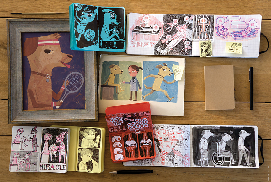
BY KATHRYN LEVY FELDMAN | Illustration by Mike Kerr
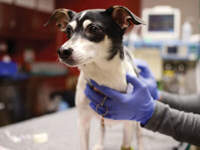
This story begins in May 2015, when Sam, my then seven-year-old golden retriever, returned from his morning walk with a decided limp. As the day progressed, he became increasingly unable to bear weight on his left-rear paw, and seemed to be in pain. A trip to the vet and an x-ray confirmed a partial tear of his left cranial cruciate ligament—important for stabilizing the stifle joint, or canine knee—and the beginning of a tear on the right side. Since the tears were not complete (which would have required surgery), I opted to try rehab with the caveat that his 95-pound weight might hinder stabilization of the joint.
Sam and I embarked on a slow but steady program of rest combined with short leash walks, weekly use of an underwater treadmill, massage, and cold laser therapy (in which beams of light are used to promote healing). He also got prescription pain medication and a non-steroidal anti-inflammatory drug. By the end of July, he was dismissed from therapy with a fluid gait and no lameness. But he was still taking high doses of meds, had trouble getting up from a lying position, and had to be helped up the stairs. In short, my seven-year-old had the mobility of a 10- or 11-year old dog.
Sam’s physical therapist suggested that he might be a candidate for a stem-cell procedure in which they would isolate his stem cells from his fat and then re-inject them into both his rear stifles. She said she’d seen incredible results, especially in large dogs suffering from osteoarthritis, which in Sam’s case had been aggravated by his injury.
In October 2015, Sam underwent stem-cell therapy in a same-day procedure. According to Thomas Masterson, vice president for business and development at Medi Vet Biologics, the originator and provider of the procedure, Sam would receive “a concentrated dose of his own regenerative cells in order to target the specific tissue by accelerating the body’s own present healing properties.” My discharge instructions were to leash-walk him for 30 days, then return for further instructions. His medication regime remained the same.
Within one week, my formerly gimpy companion, who could only get up the stairs if I pushed him from behind, greeted me at my upstairs office, wagging his tail. Within two, he was getting up and down from a lying position and covering longer and longer distances on our walks. Within three, though still on a leash, he was back at the dog park, and all my dog-walking friends were asking what I had done to Sam. After I explained the procedure, the next question was, invariably: “Why can’t they do that for me?”
I decided to find out.
Stem cells have been studied since the 1950s , when they were discovered in bone marrow. One population—hematopoietic stem cells—has been used in bone-marrow transplants for 40 years. Scientists first managed to derive stem cells from mouse embryos in 1981. But the field has really taken off in the 21st century.
In 1998, a team led by University of Wisconsin scientist James Thomson V’85 Gr’88 [“James Thomson and the Holy Grail,” Jan|Feb 2002] showed that stem cells could be grown in the laboratory from human embryos (the ethics of which was—and remains—hotly debated). Within a decade, scientists figured out how to reprogram specialized adult cells to become embryonic stem cells able to form all adult cell types, first in mice (2006) and then in humans (2007). These induced pluripotent stem cells (iPSCs) offered an alternative to embryonic stem cells and opened up the possibility that each patient could be considered a resource for the cells to treat his/her disease.
Scientists began discovering stem cells in many more of the body’s tissues and organs, including the brain, peripheral blood, blood vessels, adipose tissue, skeletal muscle, skin, deciduous teeth, hair follicle, gut, and bone. According to the National Institutes of Health (NIH), these stem cells are believed to reside in specific areas of each tissue in small numbers. Once removed, it seems that their capacity to divide may be limited, although scientists are working on ways to generate large quantities of adult stem cells in the lab and then manipulate them to generate specific cell types to be used to treat injury or disease.
Currently, blood stem cells are the only type of adult stem cells used regularly for treatment, although clinical trials are under way in the use of different types of stem cells for the treatment of Parkinson’s disease, multiple sclerosis, and Crohn’s disease, among others. To date, according to the Food and Drug Administration website, the FDA has approved “only one stem cell product, Hemacord, a cord-blood derived product manufactured by the New York Blood Center and used for specified indications in patients with disorders affecting the body’s blood forming system.”
This doesn’t mean that stem cells aren’t being used more broadly. A Google search for stem cell treatments yields a plethora of options, ranging from orthopedic to cosmetic. According to a February 2016 article in the online health-and-science publication STAT, “[a]s many as 200 stem cell clinics have cropped up in recent years, peddling injections, facelifts, and treatments for a number of devastating conditions.” These private clinics, which typically do not accept insurance for their services, “have avoided heavy regulation, in part, because they use cells extracted from a patient’s own body and because they don’t do much to those cells before re-injecting them.”
The FDA issued a public warning about stem cells in 2012, expressing its concern “that the hope that patients have for cures not yet available may leave them vulnerable to unscrupulous providers of stem cell treatments that are illegal and potentially harmful” and urging consumers to steer clear of treatments that have not been approved for use or clinical investigation by the organization.
In October 2015 the FDA issued new draft guidelines clarifying how stem cells can be used under existing regulations. They included language that cells should be “minimally manipulated,” perform the same function in recipients as they did in donors, not be combined with other materials that could raise safety concerns, and shouldn’t have a systemic effect on recipients.
These recommendations were widely perceived as representing “a crackdown on clinics offering patients experimental procedures,” according to a report in Scientific American published in September following a rare two-day public hearing (twice the usual length) on the topic at the NIH campus in Bethesda, Maryland, at which close to 90 people spoke, on all sides of the issue. “None of the FDA officials offered any reaction to the comments made at the hearing,” the article noted, “nor have they set a deadline by which they will issue final versions of the guidelines.”
Patient stories cited in the Scientific American report—which included one whose rheumatoid arthritis was in remission following stem-cell therapy and another who claimed a stem-cell injection to treat partial vision loss left him entirely blind in his affected eye—illustrate the limitations of anecdotal evidence in guiding decisions on treatment.
And the same goes, I learned, for drawing conclusions from one dog’s experience.
When I visited Penn’s Institute for Regenerative Medicine, IRM Director and Joseph Leidy Professor Kenneth S. Zaret confirmed that they were indeed “after amazing cures for illness just like what happened to Sam” by understanding how to control stem-cell fate to make them the most useful therapeutic agent—but he emphasized that thatgoal is still a long way off.
“What happened to Sam proves it’s possible,” noted Robert L. Mauck, the Mary Black Ralston Professor of Orthopedic Surgery and professor of bioengineering. “It’s up to the science to make it probable.”
George Cotsarelis, the Milton Bixler Hartzell Professor of Dermatology and chair of the department, contributed another healthy dose of scientific skepticism. “Was Sam some kind of special dog who responded to this because of his genetic makeup?” he wondered. “Are the stem cells that they injected still there? Was there some kind of natural healing going on before they even injected anything?”
Conventional medical science offers plenty of reasons to doubt whether what I did to Sam is something you should have done to your own knees, and that position isn’t likely to change in the absence of studies that are both convincing and rigorous.
“You have to do a very large study in a well-controlled way to really know what happened—and that’s the problem with a lot of the stem cell procedures,” says Cotsarelis, noting that they have yet to receive that kind of investigation.
“To really demonstrate efficacy, you have to compare placebo with the same procedure and have a blind statistical analysis of the outcome,” echoes Zaret.
It’s not that they don’t believe that it works; it’s what Zaret calls the “first step” of direct therapeutics. “I am confident in the next decade or two (and we are already seeing occasional trials) we will see more examples of direct therapeutics, but it is still going to take a while and need everybody’s support to make it work.”
In the meantime, the IRM researchers are “more focused on using stem cells and their descendents, making them into different tissue types for repair.”
There is still research being done to break down and identify the barriers that keep cells different from one another so as to figure out how to make a fully formed new kind of cell from a stem cell.
While they are doing that, however, they are also using stem cells and stem cell reprogramming technology to model human disease, developing therapeutics and diagnostics from these models. “We can now take blood from humans who have a genetic pre-disposition to liver disease or heart disease, make the cells into pluripotent stem cells, bring the cells back to liver cells that now have the genetic background that is messed up, and get more understanding of the basis for the disease as well as develop drugs to modulate those diseased cells,” says Zaret. In the laboratory they can compare “normal” liver and heart cells derived from pluripotent cells with diseased or genetically pre-dispositioned ones, as well as the effects of drugs on all of them.
Making cells “de novo” also enables scientists to develop diagnostics. In Zaret’s lab, for example, they have developed induced pluripotent stem cells from human pancreatic cancer for which there are no early diagnostic markers or cures. “You can’t even identify the patient population around which you’d want to get in there early and have early treatment because they only present late,” notes Zaret, whose mother-in-law died from the disease and inspired his research. “We figured out a way to bring the reprogrammed human pancreatic cancer stem cells back to the early stage of the disease and discovered biomarkers from that early stage,” he says. “We are now advancing those biomarkers in clinical tests to diagnose pancreatic cancer with just a blood test. We’re not at the point of presenting it to the FDA, but we are doing what we can to enable that as soon as possible.”
If Zaret is interested in the malleable properties of stem cells, Mauck and Cotsarelis are interested in their regenerative powers and how they influence inflammation and healing. Mauck’s lab makes stem-cell-based products for multiple kinds of applications that are adjuncts to existing clinical orthopedic therapies involving knee cartilage. Cotsarelis is studying how the stem cells in the hair follicle are related to wound healing, skin regeneration, and hair regeneration.
Mauck’s lab focuses on regenerating cartilage surrounding the meniscus disk in the knee. Cartilage lines the surface of the joints and has a certain thickness but no direct blood supply. That is why, when you damage your cartilage, it does not heal on its own. “There is no way to have a regenerative cell population coming in,” he explains.
Currently there are three ways doctors try to heal cartilage, none ideal.
The first is micro-fracture, which Mauck describes as “stem cell therapy although when it was invented people didn’t really understand stem cell therapy.” During the arthroscopic procedure, the surgeon creates small holes, known as micro-fractures, in the bone near the damage to release bone marrow. Stem cells from the bone marrow will fill this space and tissue forms, although it is weak, “somewhat like scar tissue,” and may only be a temporary fix. Some professional as well as recreationalathletes who have had micro-fracture are able to return to playing sports after a few months of physical therapy but, many, notes Mauck, “also perform at a lower level than they did before the procedure and their playing careers are likely shortened.”
The second method of cartilage repair is a cell-based therapy called autologous chondrocyte implantation (ACI). The surgeon scopes your knee and extracts a scoop of cartilage, which is sent to a company that separates the chondrocytes (the cells that produce cartilage) and cultures them for four to five weeks to increase the number of cells, which are then re-implanted. It takes 10 to 12 weeks before the patient can be weight bearing on the joint. This procedure is only suitable for patients who do not have cartilage damage from arthritis.
In the third procedure, called osteo-articular transfer, the surgeon harvests a small plug of healthy cartilage from areas of the joint not in critical need for weight-bearing, and transfers it to the area of damaged cartilage. The procedure is most effective for those with small amounts of damage.
All of these procedures are for those who do not need a partial or complete knee replacement, and Mauck feels they can be improved upon with the addition of products that deliver stem cells and biologics (genetically engineered proteins derived from human genes) to help stabilize and heal the repairs.
His lab, working closely with Jason Burdick, professor of bioengineering, has developed electrospun nanofiber composite materials that are fibrous networks, each with a different function, sort of “like a super-functional bandaid,” he says.
“One of the elements might be structure, another would recruit stem cells, a third might tell it to degrade a week later and become chondrocytes,” he elaborates. “We are adding biomaterials and maybe growth factors to an existing clinical scenario to help it heal quicker and better.”
These materials are in various stages of large-animal testing—a precursor to FDA approval—and yes, it takes time. “As scientists, we sometimes get stuck on mechanisms, and it’s hard to know when to stop exploring mechanisms and move a product forward.” Mauck says.
In the meantime, Mauck doesn’t think knee replacements are going to become obsolete. What are you going to replace it with? may become more of a question, however. “What I think the standard may change to is that you still might have a metal stem, but you might have a biologic top—or maybe you don’t need a metal stem and instead you have a porous metal base material and soft living material. We are modeling this in vitro currently, and our next step is to move it to animals,” he notes.
The goal of this research is to produce a product that combines stem cells and biologics and can be added to an existing procedure. “If you can work within the confines of current clinical practice, then it will be adopted more readily,” he says. “If we can build a scaffold with this new procedure that works way better than micro-fracture, then the patient who had a sporting injury in their 30s that more than likely would have [developed] devastating osteoarthritis in their 50s, now has a newer better treatment. He doesn’t get such severe erosion and doesn’t need a joint until he is 65. That would be a success.”
Cotsarelis has been researching the stem cells in hair follicles since 1988. In 2004, he made national headlines when his team discovered that implanting adult stem cells found in mice hair follicles into immune-deficient mice produced new hair follicles that produced hair [“Gazetteer,” Jan|Feb 2011]. This was huge news and suggested a future in which stem cells could be harvested from a person’s hair (before it all fell out), cultured, multiplied, and then re-injected into the scalp.
For the record, while that research does suggest that a treatment for baldness is possible, it is still a work in progress. But Cotsarelis’ research has much broader significance. “Hair follicle stem cells are actually related to wound healing and skin regeneration,” he explains. “Understanding the hair follicle really is a way of getting at all those processes.”
Stem cells in the hair follicle are unique because they are responsible for the continual regrowth of hair. This, in itself, is an unusual phenomenon because hair dies and regrows periodically during your life.
“There aren’t too many other cells in your body that do that, except for your uterus during pregnancy and that is hormonally controlled while the follicle is not,” says Cotsarelis.
He identified and isolated the stem cells in the hair follicle, which, as it turns out, were very quiescent. “They don’t proliferate very frequently, only when the follicle regenerates or if you have a wound. They will proliferate to help close a wound anywhere on your skin,” he says. Stem cells in the hair follicle are not the only stem cells that are responding to a wound, but they respond in a big way. “If there is a wound, their capacity to respond is much greater,” he adds.
This type of mass proliferating also exists in stem cells found in the cornea, which was Cotsarelis’ first area of study. His work in the field changed the way in which cornea grafts are performed in patients with chemical burns. The original protocol had been to graft corneas from cadavers that only restored vision for six months. “The reason was they weren’t bringing along the stem cells from the donor cornea,” he explains. When they did, the person would be cured. “That’s what turned me onto this field.”
For now, they are able to show that when you make a wound in the mouse it heals and regenerates fur. “Humans probably do it at a very low level,” he suggests. “In the mouse, fur is very important; if they don’t have it they don’t thermo-regulate.” In people, wounds are typically covered and closed because of infection and blood loss, perhaps at the expense of regenerating hair follicles and sweat glands and usually resulting in a scar.
Cotsarelis and his collaborators in bioengineering are trying to make hair follicles in a dish that they can introduce into a wound to stimulate healing and prevent scarring. “We haven’t really done it, but we’ve accomplished a lot of steps to get where we want to go in the development of a cell-based therapy to treat wounds,” he says. The same is true when it comes to replacing hair follicles. “We’re not there yet, but that’s the goal,” he emphasizes.
The IRM brings together experts in many fields, including cardiology, oncology, reproductive medicine, and digestive and liver diseases. Ed Morrissey, the IRM’s scientific director and professor of medicine and cell and developmental biology, for example, is doing pioneering work in cardiac repair and regeneration, specifically investigating the process by which damaged heart cells fail to proliferate due to ischemic injury, one of the major barriers to successful cardiac regeneration. Zebrafish are capable of extensive cardiac regeneration and Morrissey’s recent studies on this phenomenon, which could have a profound impact on our understanding of how to promote cardiac repair and regeneration of an adult heart, were recently published in Science Translational Medicine.
All of the work at the IRM is focused on understanding how cells work, then translate and replicate this process in the laboratory to generate and control new cells and tissue fragments and to control tissue regeneration in the body. “Our work is aimed to model and study human diseases, to develop new diagnostics and medicine, and ultimately to replace damaged, aged, or diseased body parts,” says Zaret.
To the scientists at IRM, Sam’s procedure, as successful as it may have been, is putting the cart before the horse. There haven’t been enough studies that document what it is, how it works, how long it lasts, the side effects, and how to replicate it with good odds of success.
The first randomized, double-blind, placebo-controlled, FDA-approved prospective pilot trial of the procedure that Sam underwent was conducted by David A. Upchurch, who is now an assistant professor of soft tissue surgery at Michigan State University. It has been accepted into publication by the American Journal of Veterinary Research in December 2015 although its actual publication is backlogged.
According to Upchurch, his study was the first to use the actual protocol developed by Medi Vet Biologics that Sam had, and to gain FDA approval they treated stem cells like a drug. As for conclusions, “it seems like dogs that were worse at the study onset had more of a benefit.”
Hardly conclusive. And yet as I write this in late summer, Sam is a better version of his former self. He walks two miles a day, takes half as much anti-inflammatory medication as before the procedure, and genuinely seems happy and out of pain.
“I’m just as much a patient once in a while,” admits Zaret. “I would want it in my knee, too—if I knew that it wouldn’t cause the knee to fall apart in 10 or 20 years or result in chronic pain. The threshold for success in people is different than in animals because we live a lot longer.”
Cotsarelis is blunter: “In a dog, it’s great. If it were my son, I’d tell him to have the surgery.”
A Sad Postscript

Sam passed away very suddenly in mid-September from hemangiosarcoma, a deadly cancer that invades the blood vessels. In Sam’s case it resulted in a ruptured tumor of the heart. Hemangiosarcoma is more common in dogs than in any other species and tends to develop in mid- to large-size breeds, including golden retrievers, often with no symptoms.
I was told by three veterinarians, including the one who performed the stem-cell procedure, there was no reason to believe the stem-cell treatment contributed to Sam’s demise. There is some evidence to suggest that stem cells can make existing tumors grow, but by themselves, stem cells do not cause new tumors to form.
Hemangiosarcoma is very aggressive. If Sam had the disease at the time of the treatment, he would not have survived for almost a year post-op.
While I am devastated by the loss of my faithful companion, I am comforted by the fact that the quality of his life during his last year was vastly improved by his stem-cell treatment.—KLF
Kathryn Levy Feldman LPS’09 is a frequent contributor to the Gazette.