
The greenish glow in the petri dish—a marker for the presence of germ cells—showed that veterinary-school researchers had succeeded in a decade-long quest to get male-mouse stem cells to develop into eggs.
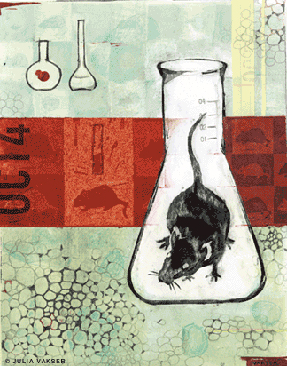
By Joan P. Capuzzi Giresi | Illustration by Julia Vakser
It was a quiet little experiment, the product of a pensive scientist’s restless imagination. A purist’s quest. But when Dr. Hans Schöler, working in his lab at the veterinary school’s New Bolton Center, transformed embryo cells from male mice into oocytes—eggs—in a petri dish, he also spawned protest, praise, and bad-pun headlines.
The religious called his work evil. The bioethicists asked if it was good for society. The skeptics said, “… but show us more.” Infertile heterosexual and gay male couples clamored for help in procuring eggs so they too might procreate.
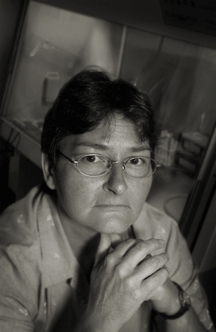
“Everyone had an interest for the sake of his or her own genetic material,” says Dr. Karin Hübner, Schöler’s research-analysis manager and lead author on the groundbreaking paper, “Derivation of Oocytes from Mouse Embryonic Stem Cells,” published in Science Online and Science Magazine in May.
Contributing to the frenzy was the mass media, which—with distorted coverage and a spate of silly headlines—seemed to ignore the fact that the experiments were performed on mice, and not humans.
But the true significance of Schöler’s work was not lost on the scientific community. Dr. John Gearhart, the C. Michael Armstrong Professor of Medicine at Johns Hopkins, whose lab was the first to isolate human embryonic germ cells, calls the discovery brilliant. “An absolute technical tour de force,” says Gearhart’s partner in the project, Dr. Peter J. Donovan, associate professor at Thomas Jefferson University’s Kimmel Cancer Center in Philadelphia. Dr. Ian Wilmut, leader of the Roslin Institute team in Scotland that cloned Dolly, the sheep, predicts Schöler’s findings are “certain to lead to a new understanding of the causes of infertility and ultimately to methods for treating some forms of infertility.”
“They have shattered another dogma,” says Dr. Jose Cibélli, Michigan State University professor of animal biotechnology and one of the founders of Massachusetts-based cloning pioneer, Advanced Cell Technology, Inc. “We knew we could make any cells from embryonic stem cells. But we would never have dreamed of having eggs produced from embryonic stem cells.”
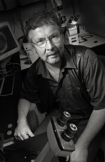
Dr. Hans Schöler: “We now have a tool to get a better understanding of the biology of the oocyte.”
It was a dream that Schöler, who runs Penn’s Center for Animal Transgenesis and Germ Cell Research, nursed for nearly a decade, starting with his co-discovery of a tiny molecule he named Oct4. Exclusive to embryonic stem (ES) and early germ—sperm or egg precursor—cells, Oct4 was described by Schöler in 1989 as a transcription factor (protein that guides DNA in encoding other proteins) that confers the hallmark property of an ES cell: pluripotency—or the ability to become almost anything. In practical terms, pluripotency is what allows that little clump of cells that forms after sperm and egg meet to spin off into neurons, heart, liver, kidney cells —and build a fetus.
After Gearhart and Donovan’s landmark harvest of human embryonic germ cells—from aborted fetuses—in 1998, researchers scrambled to induce these versatile cells to morph into every kind of cell imaginable. But they were stumped because there were two things ES cells refused to become: germ cells and the placenta-forming trophoblasts. For Schöler, now the Marion Dilley and David George Jones Chair and Professor of Reproductive Medicine in the School of Veterinary Medicine, this became a driving obsession.
Schöler sits behind his desk at the Center for Transgenesis, which consists of four major laboratories housed on two levels of the research building at New Bolton Center, Penn’s large-animal hospital, and another lab in Center City. As he searches for the right words to explain his pursuit, he seems to find speech inadequate to convey his zeal. So Schöler starts to draw. He quickly sketches the three tissue layers that form an embryo. If an embryo can produce its own germ cells to fill up its ovaries and testes, reasons Schöler, as he prods his sketch with a black marker, “why then can’t ES cells do this in a dish?”
Though he had pondered this question while still in his German homeland, he knew that it was not the place in which to seek the answer. In the persistent shadow of the Third Reich’s mission to genetically engineer a “master” race, Germany had turned a cold shoulder to human stem-cell research, strictly limiting the practice even today.
Born in Toronto, Schöler, 50, grew up in his parents’ native Germany. At the University of Heidelberg, he earned his diploma in biology followed by his Ph.D. in molecular biology. And he developed a keen interest in gene regulation and developmental biology, particularly as they relate to the germ line.
“I’ve always been interested in what distinguishes body cells from germ cells,” he says. “Germ cells are fascinating because they link one generation to the next.”
After several years in academe and industry, Schöler went to the European Molecular Biology Laboratory (EMBL), to head his own research group in 1991. There, he was rejoined by former colleague Karin Hübner.
Hübner, who grew up on skis and surrounded by animals in the Bavarian Alps, had always had a knack for science. She studied biochemistry, became certified as a laboratory technician, and went to work in the lab of Nobel laureate Feodor Lynen, who was investigating the regulation of cholesterol and fatty-acid metabolism. Hübner then moved on to the fields of cell biology and neurochemistry, and finally molecular biology. She worked with Schöler on the Oct4 protein and shared his gusto for the mysteries and promises of ES cells.
By the late 1990s, Schöler, who currently sits on the stem-cell research advisory board for Germany’s conservative Christian Democratic Party, had achieved fame for his accomplishments in the stem-cell field. But with little enthusiasm or funding for his research in Germany, he set his sights on the University of Pennsylvania. Philadelphia was emerging as the international hub of stem-cell research, and Penn offered a sturdy emphasis on the basic sciences.
“Penn has more talent in its clinical and basic-science labs than anyone else in the world,” says Dr. Glenn McGee, assistant professor of medical ethics at Penn’s School of Medicine. “No other institution can compete with Penn when it comes to answering the question, ‘What’s in the dish?’”
Most importantly, says Schöler, Penn offered Dr. Ralph L. Brinster V’60 Gr’64, the Richard King Mellon Professor of Reproductive Physiology at the veterinary school. Brinster, whom Schöler calls “the godfather of so many things,” is perhaps the world’s premier contributor to the field of genetic modification of the germ line. Schöler welcomed the opportunity to collaborate with him. So in the summer of 1999, he moved with his wife—an attorney—and two school-age sons to the Philadelphia suburbs.
“Hans Schöler’s arrival really strengthened stem-cell biology at Penn,” says Narayan Avadhani, the Harriet Ellison Woodward Professor of Biochemistry and chair of the animal-biology department at the veterinary school. Avadhani says that the Brinster-Schöler stem-cell tag team propels the veterinary school —which, according to officials in its dean’s office, has invested more than $15 million on stem-cell research thus far—ahead of other stem-cell powerhouses like Johns Hopkins and the University of Wisconsin to become the world leader in the field.
Hübner, who had spent some time working in Texas, was eager to bring her ES work to the States. She and a few others followed Schöler and, by late summer, Hübner was hooked into a new mission—to generate oocytes from mouse ES cells. Though enticed by the idea of pushing ES cells to make this transformation, a notion she and Schöler had discussed for several years, she was not convinced that her microscopic subjects would cooperate.
“I can’t say I figured, ‘Yes, yes, they’re going to do it,’” she recalls. “It was more like, ‘Let’s try it, and let’s see.’”
The key to the project was finding a way to recognize the germ cells, if and when they developed. To this end, Schöler and Hübner had years earlier created a stem-cell marker using green fluorescent protein (GFP). As soon as she began the current endeavor, Hübner transformed her ES line—derived from male-mouse embryos—with a tailored Oct4 transgene designed to activate GFP only in early germ cells. Hence, cells in the early germ stage were programmed to glow green.
For months, Hübner toyed with the construct, altering the Oct4 and manipulating the cell densities and culture conditions.
Then came the “eureka” moment. On that February afternoon, as Hübner peered into her microscope, her eyes met the faint glint of green. Germ cells.
“It was like, ‘Whoa, they’re green green!’” Hübner remembers.
Green as they were, they were scant in number. So few that Hübner wondered whether they really were germ cells. But when she checked in on them the next day, she discovered more cells that had cropped up overnight: “I started photographing like crazy and then I ran to Dr. Schöler’s office and said, ‘I think we’ve got them.’ Then everyone started running over to the scope to have a look.”
Over the next few weeks, Hübner ran tests to verify that what she was seeing were germ cells. Then she optimized her culture formula. The picture that emerged was that of a dynamic, ovary-like microenvironment in a dish, where germ cells could mature into oocytes and, in some cases, embryoid structures.
“We were fascinated to see that the mouse cells were capable of producing oocytes that recruit adjacent cells to surround and nurture eggs so structures similar to the natural follicles can form,” Schöler says. “But even more amazing was that the morphological changes were paralleled by physiological function. Proteins and hormones were expressed in a coordinated fashion pretty similar to that of an estrous cycle. The oocytes could even enter meiosis and eventually develop into embryos.”
Stem-cell pioneer Gearhart views this as an excellent teaching model from which to observe how eggs are made. “We may now be able to study oogenesis in a dish,” he exclaims.
By culture Day 8, the Science paper reports, some 40 percent of the plated cells had turned green, indicating early germ cells. GFP expression declined slightly over the next four days as the cells began to separate from each other, similar to germ-cell migration in vivo. ES cell aggregates appeared to behave like follicles, flanking the presumed germ cells and secreting an estrogen precursor. Evidence of meiosis—reproductive cell division—was present after 16 days in culture. Around Day 26, apparent oocytes that had attained the size range of natural, mature oocytes (50-70 microns) were released from their supporting cells, much as normal eggs are released from their follicles during ovulation.
Perhaps most surprising to Schöler and his colleagues was the emergence, at about Day 43, of structures similar to mouse preimplantation embryos. Schöler believes these embryoid structures are parthenotes, embryos that develop from unfertilized eggs. The team also observed several structures that resembled blastocyst-stage embryos, the source for ES cells.
In her modest-sized lab, Hübner surveys a group of plates that sit in two large incubators, and she ponders the demands the cells place on her. “Even if it’s a weekend, the cells don’t know,” she says. “They still get hungry and need to be taken care of. That can get annoying.”
Sometimes Hübner works at the incubator hood for six straight hours, followed by another six at the microscope. Despite such tedium, she loves her work. “To see these events going on in the cell nucleus under a scope is one of the most beautiful things there is,” says Hübner, 46, who is invigorated by the chance to peer in on these units that represent the smallest parts of our bodies. “It’s like getting to know yourself. These cells change all the time, and one realizes when they are happy and when they are not happy.”
Hübner removes a culture plate the size of a hockey puck from one of the incubators. It contains a 26-day culture, and she places it under a souped-up microscope that, she says, cost $130,000. The scope, a Leica, is hooked up to a computer with a monitor that facilitates visualization of the green fluorescence. As she adjusts the wavelength, a band of hazy green—presumably germ cells—shines under the scope.
The green streak, which flanks a denser cluster of support cells that appear grayish, looks moth-eaten. Cell-to-cell contact is critical for germ-cell maturation, Hübner explains. But, as in the body, germ cells maturing in vitro suffer massive die-offs and the survivors gradually lose their tight adherence. Once the remaining oocyte-like structures in the dish are released from their follicular scaffolding, their pursuit of independence causes them to scatter.
Their moth-eaten appearance—easily confused with a failed culture—is probably responsible for the anonymity that cultured eggs seem to have had thus far, Hübner explains: “Morphologically, it does not look attractive because lots of cells are dying. People have told me, ‘I think I’ve seen these cells in the dish and thrown them away.’”
Were it not for the green GFP marker, Schöler and his colleagues might have done the same. Schöler always knew he would one day culture oocytes. It was a conviction that, as far back as a decade ago, he shared with those who inquired about his plans for the GFP he and Hübner were developing.
“They would say to me, ‘Ah, that will never work,’” Schöler recalls.
Although it seems to have worked, the naysayers have not been silenced. “Now,” he chuckles, “they’re saying, ‘Let’s see if these eggs can be fertilized.’”
These days, Hübner is busy refining her culture method in order to improve the cell yield from the current 20-25 oocytes per plate. Her goal is to generate a continuous supply of germ cells in different phases of maturation. The team can then investigate whether the cells they have cultured are truly eggs and, if so, viable ones. Her plans include differentiating the cultured cells, manipulating the culture conditions, performing chromosomal analyses of supposed oocytes, observing meiotic divisions, and attempting fertilizations and implantations.
Schöler et al. are also considering trying to harvest sperm from ES cells in the dish. However, Schöler says, such attempts will likely be more difficult. The reason his ES cells, though male in origin, generate eggs rather than sperm is that production of the latter requires far more complex signaling. “Cells need to relax in a more perfect way in order to become sperm,” he explains. Hence, oocytes are produced as default products.
Much of the early media coverage of Schöler’s research raised false hopes of eggs-for-all. On May 2, the day after the article was published in Science Online, The Washington Post put an interesting slant on the team’s research. Basing their assertion on the fact that eggs were generated from male ES cells, the Postsuggested that gay couples were now one step closer to procreating together. The European media were just as guilty: They frenzied readers with headlines like, “Eggs Over Easy” and “Who Needs Ovaries?”
Schöler says the onslaught of phone calls was immediate. In the weeks after the paper was published, he fielded hundreds of inquiries from around the world. Senators and governors phoned in and e-mailed, as did infertility clinics, anti-gay organizations, laboratories seeking to collaborate, and hopeful study participants.
But while mice are good models for the study of human physiology, these findings cannot necessarily be extrapolated to people. “Often what works in mice doesn’t even work in rats,” Donovan says.
Nevertheless, bioethicists are preparing for the worst, or—depending on the individual’s position—the best. One group praises Schöler’s research (U.S. patent pending) as setting the stage for potential egg factories that would take the pressure off women to donate. Many medical researchers, like Cibelli, embrace this position. “As a cloner,” he says, “I can say the sex appeal is having an unlimited supply of eggs without having to worry about donors.”
The other group of bioethicists, says Schöler, denounces the research. “They say, ‘Now you’re actually creating life in a dish.’ But some of the things they see as big problems are actually not. They think therapeutic cloning is a slippery slope toward reproductive cloning, which we know is not feasible in humans.”
Penn’s McGee describes stem-cell ethics as a highly charged field that touches on hot buttons like abortion, fetal-tissue use, cloning, organ transplantation, gene therapy, and animal welfare. And it challenges society to do the impossible—agree on a uniform definition of the word embryo. McGee, who contends that one cannot use the word unless one thinks it can be born, recently asked 30 embryologists to define an embryo. He got 30 different answers. But characterizing an embryo may well be simpler than ruling on whether or not it can be killed.
If the germ cells prove to be viable enough for nuclear transfer (cloning) but not for producing offspring, then Schöler’s work may deflate the main argument against therapeutic cloning—that it manufactures embryos so that they can be destroyed.
“We have in this research a real opportunity to possibly define something that is not an embryo but is like one,” McGee says.
Conspiracy theorists have weighed in on the argument, claiming that the manufacture of eggs in a dish could really be a cloaked attempt to ultimately take women out of the reproduction picture. (Never mind the fact that scientists have not yet come up with a way to grow a uterus in a dish.)
While some fret over remote possibilities, Dr. Ruth Hubbard, professor emeritus of biology at Harvard and a founding member of the Council for Responsible Genetics, preaches reserve: “Long-term scenarios are really not very useful.”
But one thing is for sure, says Dr. Rosemarie Tong, distinguished professor of healthcare ethics at the University of North Carolina in Charlotte. “We need to let the ethical-social-legal-nexus catch up to these complexities.”
The U.S. government has hardly rushed to support human ES-cell research. Countries like Sweden, Israel, Singapore, and Australia have comparatively fewer restrictions.
Of the $27 billion National Institutes of Health (NIH) budget for fiscal 2003, $419 million has been allocated for stem cell research. (Schöler holds a $1.25 million NIH grant to study the role of Oct4 during germline development.) But President George W. Bush, who has articulated his ethical concerns about stem-cell research, limited federal funding to the 70 or so stem-cell lines created on or before August 9, 2001 (of which only 12 are considered potentially viable for research). Some critics decry this as a possible conflict of interest: Secretary of Health and Human Services Tommy Thompson is the former governor of Wisconsin; the largest stem-cell patent holder in the country, the University of Wisconsin Alumni Research Foundation (WARF), holds the patents on some 60 of the approved lines.
In addition, more than 30 states have laws on the books pertaining to stem-cell research. In Pennsylvania, the Abortion Control Act of 1989 is somewhat ambiguous on the issue. Although the Rendell administration has not yet clarified its position, the state’s attorney general supports President Bush’s policy, says Sean Connolly, spokesman for Attorney General Mike Fisher.
Schöler says the federal restrictions on the use of ES cells could hamper advances in therapeutic cloning. “Although I know that we still have a long way to go and maybe all attempts at the end may turn out to be futile, I have a problem with people trying to tell me I can’t save my beloved if I have the ability to do it,” he says.
While McGee believes that “the future of medical care probably won’t involve extracting goo from embryonic stem cells,” he says stem cells teach us more about our bodies’ cellular mechanisms so we might one day develop pharmaceuticals designed to make our own cells work better.
It is too early to gauge the impact stem cells will have on the future of medicine. Other newer medical technologies like gene therapy and somatic (mature) cell cloning have fallen shy of their promises. Since application is everything in science, history will likely judge the value in harvesting eggs from ES cells in light of what it ultimately does for humanity. But what of science for the sake of science?
Schöler, the pensive purist, acknowledges that it “would be wonderful if our finding paved the way to therapies, as many people hope. But even if this turned out not to be the case, I think we now have a tool to get a better understanding of the biology of the oocyte, the one cell that I think is the most amazing of all. At the end of my life, I hope to have caught at least a glimpse of its magic.”
Joan P. Capuzzi Giresi C’86 V’98 wrote about her experience studying at Penn’s School of Veterinary Medicine in “Alumni Voices: The Deluxe Edition” in November/December 2002.
